Your cart is currently empty!
Researchers Discover Preserved Blood Vessels in T. Rex Fossil — Here’s How They Survived Millions of Years

When Jerit Mitchell stared at the computer screen displaying scan images from a 66-million-year-old bone, he knew something was wrong. The structures threading through the fossilized rib didn’t match anything he’d seen in paleontology textbooks. His mentors gathered around the monitor, their expressions shifting from curiosity to excitement as they realized what they might be looking at.
Contents
show
Six years of rigorous research would follow that initial moment of discovery. What began as an undergraduate physics student’s routine examination of fossil scans would evolve into one of the most significant paleontology breakthroughs in recent years. Hidden within the bones of history’s largest predator lay evidence that would challenge fundamental assumptions about what can survive across deep time.
The implications of this discovery extend far beyond a single fossil. What scientists found preserved inside Scotty the T. rex could revolutionize our understanding of dinosaur biology and open new frontiers in the search for ancient life’s secrets.
Incredible Discovery Inside World’s Largest T. Rex Fossil

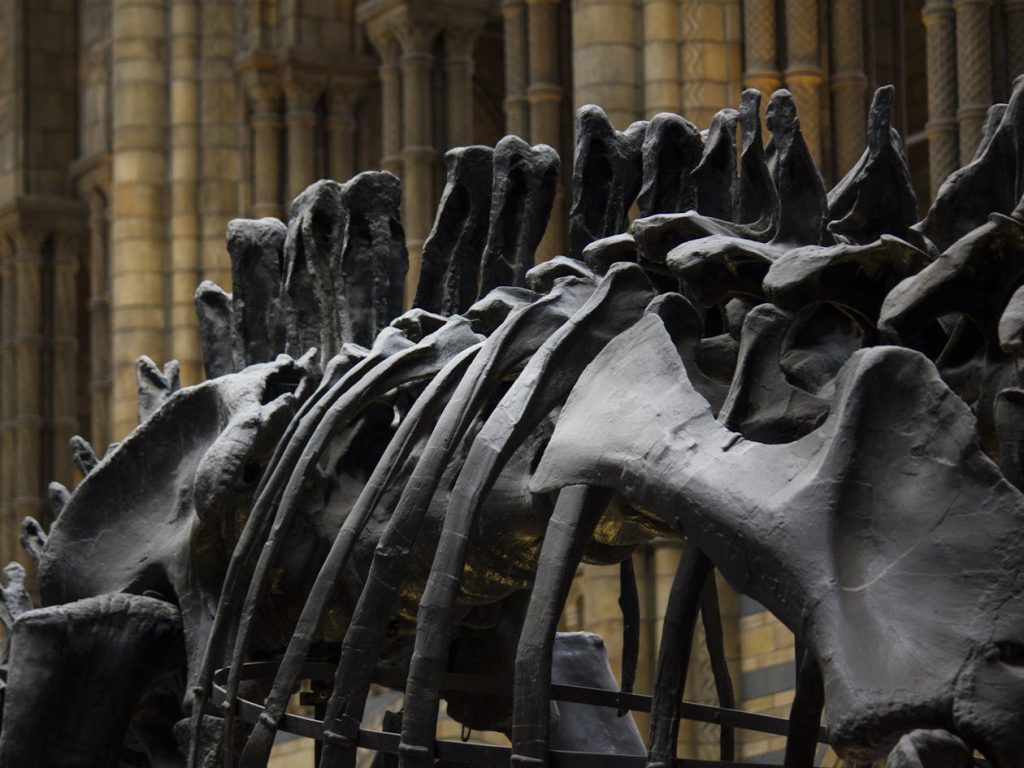
Scientists have discovered preserved blood vessels inside Scotty, the largest Tyrannosaurus rex fossil ever found. This exceptional specimen, housed at the Royal Saskatchewan Museum in Canada, represents one of the most complete T. rex skeletons ever recovered from the Late Cretaceous period.
Discovered in 1991 near Eastend, Saskatchewan, Scotty would have weighed nearly 20,000 pounds when it roamed ancient river plains 66 million years ago. The fossil’s remarkable preservation stems from specific environmental conditions that occurred during burial in the Frenchman Formation.
With 65% of its skeleton recovered, Scotty provides paleontologists with an unprecedented window into T. rex anatomy and behavior. The specimen’s completeness allows researchers to study not just individual bones, but entire biological systems that operated within this apex predator.
The discovery represents a significant advancement in soft tissue paleontology, a field that has gained momentum as sophisticated analytical techniques become available to researchers. Such findings were considered impossible just decades ago.
Published results in the journal Scientific Reports document the first detailed analysis of large angiogenic blood vessels in dinosaur fossils, providing new insights into how these ancient animals healed from injuries.
Student’s Routine Scan Reveals Extraordinary Hidden Structures
Jerit Mitchell first encountered the preserved blood vessels in 2019 while working as an undergraduate physics student at the University of Regina. His background in particle accelerator technology proved crucial for applying advanced analytical methods to fossil research.
“Normally, what gets preserved in the fossil record is only just the hard parts—just the bones or the teeth,” Mitchell explained about typical fossilization processes. His discovery challenged conventional wisdom about what biological materials can survive fossilization.
The initial finding occurred during the routine examination of computed tomography scans of Scotty’s rib bones. What appeared as anomalous structures on the imaging data prompted closer investigation by the research team.
Six years of subsequent research involved multiple analytical techniques and collaboration between physics and paleontology departments. Mitchell’s journey from undergraduate discovery to doctoral research demonstrates how interdisciplinary approaches can yield breakthrough scientific results.
The research team employed cutting-edge synchrotron radiation techniques available at only select particle accelerator facilities worldwide. This technology enabled non-destructive analysis of the fossil’s internal structures with unprecedented detail.
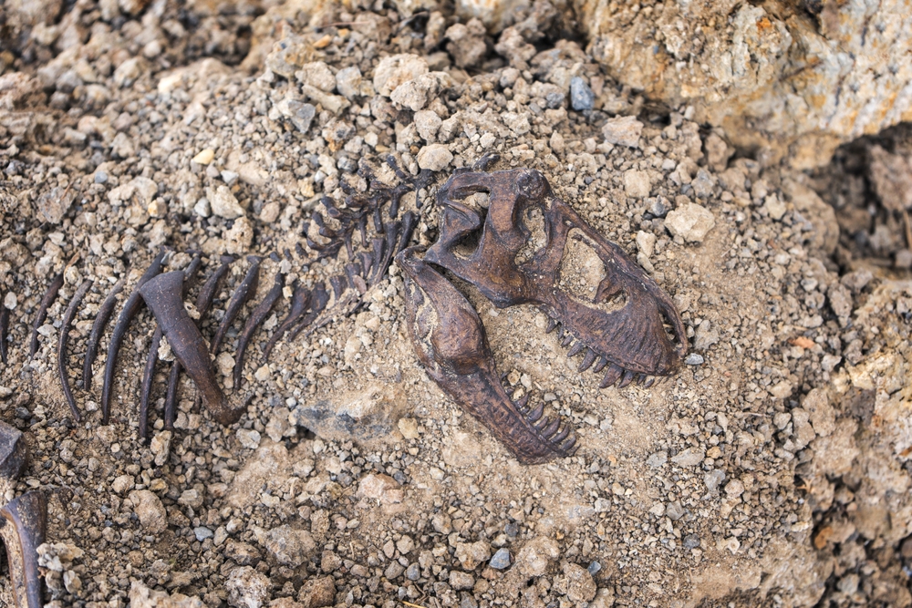
Scotty’s Violent Life Recorded in Battle-Scarred Bones
Evidence preserved in Scotty’s skeleton suggests the massive predator lived a particularly violent existence during the Late Cretaceous period. Multiple injuries documented throughout the fossil indicate frequent combat with other dinosaurs or environmental hazards.
Paleontologists have identified numerous pathologies, including fractures on the tail, skull, and rib, that provide insight into T. rex behavior and survival capabilities. These injuries likely resulted from territorial disputes or competition for mates among adult tyrannosaurs.
One rib bone displayed a large fracture that had partially healed before the animal’s death. This incomplete healing process created the specific conditions necessary for blood vessel preservation within the bone tissue.
The healing fracture indicates Scotty survived for several months after sustaining the injury, demonstrating the remarkable resilience of these apex predators. Such survival capability suggests sophisticated physiological mechanisms for recovery from trauma.
Bite marks and other evidence of combat found throughout tyrannosaur fossil records support theories about aggressive behavior among mature individuals competing for resources and territory.
Revolutionary Synchrotron Technology Peers Inside Ancient Bones
Advanced synchrotron X-ray imaging allowed researchers to examine the fossil’s interior without causing any damage to the specimen. This non-destructive approach preserves invaluable fossils for future research while enabling detailed current analysis.
Conventional medical CT scanners lack sufficient power to penetrate the dense, mineralized bone that results from millions of years of fossilization. Synchrotron facilities generate high-intensity X-rays capable of imaging even the densest geological materials.
Particle accelerators produce synchrotron light by accelerating charged particles to nearly the speed of light, then directing them through magnetic fields. The resulting radiation provides researchers with exceptionally detailed imaging capabilities.
“But we can actually have the soft tissues preserved in rare circumstances, and these can tell us a lot more about how dinosaurs lived millions of years ago,” Mitchell noted about the potential for soft tissue discovery in fossils.
Three-dimensional reconstruction techniques enabled researchers to create detailed models of the preserved vascular structures, revealing branching patterns and size distributions that characterize blood vessel networks.
Healing Process Creates Perfect Preservation Conditions
Bone fractures trigger massive increases in blood vessel activity as part of the natural healing process. This angiogenesis brings nutrients and immune cells to damaged areas, facilitating tissue repair and regeneration.
Scotty’s incomplete healing created an extensive network of blood vessels that became preserved through specific mineralization processes. The timing of death during active healing provided optimal conditions for soft tissue fossilization.
“Preserved blood vessel structures, like we have found in Scotty’s rib bone, appear linked to areas where the bone was healing,” study co-author Mauricio Barbi explained about the connection between injury and preservation.
Enhanced blood flow during healing increases the concentration of iron-rich blood in affected areas. This iron content becomes crucial for the chemical processes that enable long-term preservation of organic structures.
The research suggests that targeting fossils with evidence of injury or disease may increase the chances of discovering preserved soft tissues, providing new strategies for paleontological exploration.
Iron-Rich Mineralization Preserves Organic Structures
Blood vessels survived fossilization through iron-rich mineralization that created durable casts of the original organic structures. Chemical analysis revealed these vessels were preserved as iron oxide minerals, including goethite and hematite.
The preservation process involved two distinct layers of mineral formation, reflecting the complex environmental history that led to Scotty’s exceptional fossilization. Initial pyrite formation was later partially oxidized to create the final mineral composition.
Iron concentration in the preserved vessels reached levels multiple orders of magnitude higher than surrounding bone matrix, confirming the biological origin of these structures rather than random mineral formation.
Synchrotron X-ray fluorescence mapping revealed the specific chemical composition of the preserved vessels, including traces of manganese and other elements associated with blood chemistry.
The iron-based preservation mechanism aligns with proposed Fenton reaction pathways, where iron from blood hemoglobin creates cross-links in protein structures that stabilize them over geological time periods.
Soft Tissue Preservation Challenges Traditional Fossil Expectations
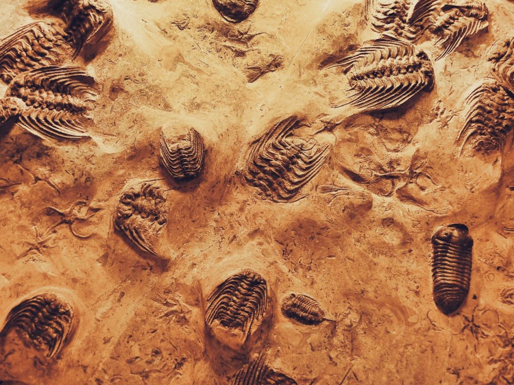
Soft tissue preservation in fossils represents an extremely rare phenomenon that provides much richer information about ancient life than bones alone. Examples include preserved muscles, ligaments, pigments, and skin impressions that reveal details about dinosaur appearance and behavior.
Previous claims of dinosaur blood cells and other soft tissues have faced scientific scrutiny, with some interpretations later revised as mineral formations rather than biological remains. Rigorous analytical methods are essential for validating genuine soft tissue discoveries.
The current research employed multiple complementary analytical techniques to confirm the biological origin of the preserved structures. This multi-method approach reduces the likelihood of misinterpreting mineral formations as biological remains.
Advances in analytical technology have enabled the detection of increasingly subtle traces of ancient biology that were previously undetectable. These capabilities continue expanding as new techniques become available to paleontologists.
The rarity of soft tissue preservation makes each confirmed discovery particularly valuable for understanding extinct organisms and the fossilization process itself.
3D Models Reveal Extensive Vascular Networks
Three-dimensional reconstruction of the preserved blood vessels revealed intricate branching patterns characteristic of angiogenic networks formed during bone healing. Vessel diameters ranged from 100 to 500 micrometers, consistent with blood vessels that supply healing bone tissue.
Some vessel segments appeared completely mineralized while others remained hollow, creating a complex three-dimensional structure that could be segmented and analyzed using specialized software. The preserved networks extended from the bone’s interior medullary cavity to its outer cortical regions.
Larger vessels measuring up to 1 millimeter in diameter were identified near the fracture site, representing major blood supplies that would have carried substantial blood flow to healing tissues. These dimensions exceed typical Haversian canal sizes found in normal bone.
The vascular architecture resembles healing patterns observed in modern animals, suggesting similar physiological responses to injury across vast evolutionary timescales. Such comparisons provide insight into the evolutionary conservation of healing mechanisms.
3D printing technology enabled the creation of physical models from the digital reconstructions, allowing researchers to examine the preserved structures from multiple angles and share findings with scientific colleagues.
Chemical Fingerprints Confirm Biological Origin
X-ray fluorescence mapping revealed distinct chemical compositions that distinguish the preserved blood vessels from surrounding bone matrix and random mineral inclusions. Iron and manganese dominated vessel chemistry, while calcium characterized normal bone areas.
No similar vascular structures were found in healthy bone sections from the same fossil, supporting the hypothesis that preservation relates specifically to injury-induced angiogenesis. This selectivity strengthens arguments for biological rather than purely geological origins.
Energy-dispersive spectroscopy and X-ray absorption spectroscopy provided detailed chemical analysis confirming the iron-oxide composition of vessel walls. These techniques revealed specific mineral phases, including pyrite, goethite, and hematite.
Chemical microprobing at multiple locations within individual vessels showed consistent compositions that match expected preservation pathways for iron-rich biological tissues. Such consistency argues against random mineral formation.
Trace elements, including lead, nickel, and zinc, were detected within the vessels, potentially representing original biological chemistry or environmental contamination during fossilization.
Discovery Opens New Frontiers in Dinosaur Biology Research
The blood vessel discovery suggests new research directions for understanding dinosaur physiology and healing capabilities. Future studies may compare vascular structures across different dinosaur species to trace evolutionary changes in healing mechanisms.
Comparative analysis with modern bird and crocodile healing could provide insights into the evolutionary history of vertebrate repair mechanisms. Such comparisons may reveal which healing features are ancient versus recently evolved.
The research methodology developed for this study can be applied to other fossils showing signs of injury or disease. This targeted approach may increase success rates for discovering additional soft tissue preservation.
Cross-disciplinary collaboration between physics and paleontology proved essential for this discovery, demonstrating the value of applying advanced analytical techniques to traditional paleontological specimens.
Access to multiple synchrotron beamlines enabled comprehensive chemical and structural analysis that would not have been possible with single-technique approaches.
Technology Advancement Transforms Paleontology Methods
Synchrotron radiation facilities represent cutting-edge technology that is revolutionizing paleontological research methods. These particle accelerator installations provide analytical capabilities unavailable through conventional laboratory equipment.
Non-destructive analysis techniques preserve valuable fossil specimens while enabling detailed internal examination. This approach allows repeated analysis as new methods become available without damaging irreplaceable materials.
Advanced imaging software enables three-dimensional reconstruction and analysis of complex internal structures. Such visualization capabilities were impossible with traditional two-dimensional sectioning methods.
Multiple complementary analytical techniques verify results and reduce risks of misinterpretation. This multi-method approach has become standard practice for controversial findings in paleontology.
The integration of physics-based analytical methods with paleontological research represents a growing trend toward interdisciplinary approaches in fossil studies. Such collaboration between fields continues expanding the boundaries of what can be learned from ancient remains.