
Your cart is currently empty!
New Bone Regeneration Technology Could Reduce Painful Graft Surgeries

For some children born with cleft lip and palate, repairing a gap in the jaw can mean taking healthy bone from their own hip and moving it somewhere else. Researchers in Australia have now developed a tiny biodegradable material that could eventually encourage the body to rebuild that missing bone itself.
Contents
show
The early results are promising, but they come with an important limitation: the material has only been tested in animals so far. Even so, researchers found roughly 80% more new bone in treated rats after eight weeks, raising the possibility of a less invasive approach to a surgery children have faced for decades.
The Jaw Surgery Children May Wait Years For
Cleft lip and palate affects around 1 in 700 children, according to the research team. While the visible cleft is often repaired during infancy, the underlying bone defect can require a different procedure later, particularly when children need enough bone to support developing teeth.
Many children with larger defects wait until they are around 10 to 12 years old before surgeons can perform a bone graft.
Associate Professor Chun Xu, a Sydney Horizon Fellow at the University of Sydney, described repairing this bone defect as one of the biggest challenges facing children born with cleft lip and palate.
“One of the biggest challenges for children born with cleft lip and palate is repairing the bone defect in the jaw,” Xu said. The procedure can eventually provide the missing bone, but getting there requires another significant operation.
Why The Bone Comes From The Hip

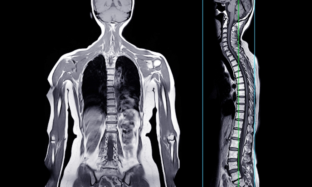
The standard technique is known as an autograft, which means surgeons take bone from another part of the patient’s own body. A common source is the iliac crest near the hip, where bone can be harvested and transferred into the jaw defect.
The approach has an obvious advantage because the patient’s own bone is being used. However, it also creates a second surgical site, meaning the child has another wound to recover from in addition to the original procedure.
“Our long-term goal is to develop materials that help the body regenerate bone naturally and reduce the need for these invasive and painful procedures,” Xu said. The new nanomaterial is designed around precisely that idea.
The Nanomaterial Turns On A Natural Repair Signal
Instead of supplying the body with large amounts of manufactured growth factors, Xu’s team looked at signals that are already present in the body. One of those signals is TGF-beta1, a protein involved in tissue repair that can remain in a latent, inactive state until it receives the right chemical cue.
The researchers developed calcium-aluminosilicate nanoparticles containing microscopic pores. These particles create a small alkaline environment around themselves, which can activate the dormant protein and help trigger the body’s bone-building response.
According to the published research described in the source, the material activated TGF-beta1 at more than 10 times the efficiency of a conventional alkaline approach. Xu explained that the team was trying to make use of repair mechanisms that already exist inside the body rather than relying entirely on externally supplied signals.
“Our body already contains many of the signals needed for tissue repair. We’ve developed a material that can help activate those signals at the right place and time,” Xu said. Once activated, the signal can attract bone-forming stem cells toward the damaged area.
The Material Is Designed To Disappear
The particles are intended to break down gradually as new bone forms. If the approach works as designed, the artificial material would eventually be replaced by tissue produced by the patient’s own body.
That makes the concept particularly interesting for reconstruction because the goal is not simply to fill a hole permanently. Instead, the material is being used as a temporary trigger that helps the body’s own cells rebuild the missing structure.
Rats Produced Around 80% More New Bone
The researchers tested the material using a critical-size defect in a rat skull. This type of experiment creates a bone defect large enough that it is not expected to heal on its own, allowing researchers to measure whether a treatment actually improves regeneration.
After eight weeks, the rats treated with the nanomaterial had roughly 80% more new bone than animals given a control material. The result provides evidence that the particles can stimulate substantial bone formation under controlled experimental conditions.
The finding is significant for the research team, but it does not establish that the same result will occur in humans. Animal studies are an early stage of medical development, and additional testing is needed before the material can be considered for human treatment.
The Particles Also Help Blood Clot
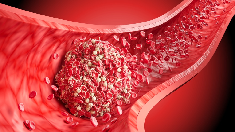
The material has another feature that could be useful during surgery because it promotes blood clotting in around 30 seconds. Rapid clot formation can help stabilize a scaffold at the surgical site while the early stages of healing begin.
The researchers describe the particles as combining clot-promoting and bone-building functions in a single platform. Keeping the material stable at the repair site could be particularly useful because a scaffold that moves before new tissue forms may not support the intended reconstruction.
The Potential Uses Go Beyond Cleft Palate Surgery
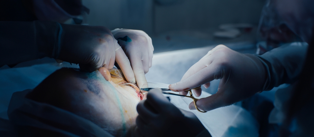
The research began with a specific problem, but the underlying challenge appears in many areas of medicine. Large bone defects can occur after serious injuries, tumor removal, tooth loss and other reconstructive procedures.
The source article reports that more than four million bone repair procedures are carried out worldwide each year. If future studies show that this technology is safe and effective in people, researchers could eventually investigate whether the same principle works for other types of bone repair.
Potential applications could include:
- Serious injuries: Large gaps left after accidents could potentially be supported with regenerative scaffolds.
- Tumor removal: Removing diseased bone can leave defects that require reconstruction.
- Dental procedures: Some patients need additional bone before certain dental implants can be placed.
- Facial reconstruction: Materials tailored to individual defects could potentially support complex jaw repairs.
Those possibilities remain hypothetical at this stage. The researchers first need to establish whether the material can safely regenerate bone in humans and whether the new tissue remains strong over time.
Human Treatment Is Still A Long Way Off

The biggest caveat is straightforward: no human patients have received the nanomaterial. The University of Sydney says additional testing will be needed before human trials can begin, so the current findings should be viewed as preclinical research rather than a new treatment option.
There are also specific questions surrounding children with developing jaws. Researchers will need to understand how quickly the material breaks down, what amount should be used and how the newly formed bone behaves as a child grows.
The location of the repair creates another concern because developing teeth sit close to the jaw defect. A material designed to activate biological repair signals must be studied carefully to determine whether it could have unintended effects on surrounding tissues.
Why The Rat Results Still Matter
The limitations do not erase the value of the experiment. A critical-size defect is deliberately difficult to heal, so producing substantially more bone than the control group gives researchers evidence that the material is doing something biologically meaningful.
The next steps will determine whether that effect can be reproduced safely in larger studies and eventually in people. Moving from a successful animal experiment to a reliable human treatment is a lengthy process, particularly when the potential patients are children.
A 3D-Printed Scaffold Could Be Next
Xu’s team is also looking at how the technology could be combined with advanced 3D printing. That could eventually allow a scaffold to be shaped around an individual patient’s specific jaw defect rather than relying on a standard design.
“Every patient is different and every bone defect is different. In the future, we hope to combine these materials with advanced 3D-printing technologies so treatments can be tailored to the specific needs of each patient,” Xu said.
The vision is still experimental, but it points toward a very different approach to bone reconstruction. Instead of removing healthy bone from a child’s hip and transferring it to the jaw, doctors could one day place a temporary scaffold that helps the child’s own cells rebuild the missing structure.
For now, the nanobone remains an experimental technology backed by animal data rather than a procedure available to patients. But if future studies can answer the safety questions, the idea of repairing a child’s jaw without taking bone from another part of their body could move from laboratory research toward clinical reality.