Your cart is currently empty!
Rare Surgery Helps Man See Again After Losing Vision for Twenty Years Using His Own Tooth

For more than two decades, Brent Chapman lived in total darkness, relying on memory, sound, and touch to navigate the world around him. A rare allergic reaction when he was just 13 years old had left him blind, plunging him into a world where daily life was shaped by uncertainty and loss. Doctors tried again and again to restore his vision through corneal transplants and other procedures, but each one failed, forcing Chapman to adapt to a reality without sight. Over the years, he adjusted to his condition, but the longing to see again, to make eye contact, to recognize faces, to experience the vivid details of the world never went away.
Contents
show
Now, at 34, Chapman’s life has been transformed in the most extraordinary and almost science fiction-like way: surgeons implanted one of his own teeth into his eye, restoring his ability to see. The procedure, known as osteo-odonto-keratoprosthesis (OOKP), is incredibly rare and has only been performed a few hundred times worldwide since its development in the 1960s. For Chapman, it was a last-resort option after years of unsuccessful treatments, and the result was nothing short of miraculous. Today, he has functional vision in his right eye, something he never thought he would experience again and he’s sharing his story of how a tooth became the key to unlocking sight.
The Long Road From Blindness to Hope
When Chapman first lost his sight, it was the result of Stevens-Johnson syndrome, a rare autoimmune reaction triggered by a dose of ibuprofen. This sudden reaction severely damaged the surface of his eyes, leaving him blind in both. As a teenager, the transition from sighted life to blindness was overwhelming. His family recalls how quickly their lives changed from everyday activities like playing basketball and biking around the neighborhood to the slow, deliberate process of relearning independence through mobility aids, tactile markers, and memory.
Over the years, Chapman pursued every option doctors suggested. Corneal transplants, which are typically the first line of defense for damaged corneas, were attempted multiple times. Each time, his sight returned briefly, only to fade away as the transplant failed to heal properly. The repeated disappointment was crushing.
Chapman has said that every glimpse of vision he regained made losing it again that much harder, sending him into cycles of hope and despair. “It was very devastating when I would lose that vision again, so we couldn’t keep going down that road,” he recalled. By the time he reached adulthood, Chapman had undergone more than 50 surgeries, but still lived in total darkness.
The toll reached beyond medicine, touching people’s emotions and social lives. Chapman has spoken about the depression he felt as the world remained hidden from him, the frustration of losing his independence, and the grief of missing out on visual milestones others take for granted. He found ways to cope, working as a massage therapist and building a fulfilling life, but the desire for vision was always present. That’s why, when Dr. Greg Moloney suggested the radical “tooth-in-eye” surgery, Chapman’s initial skepticism soon gave way to hope. It was risky, experimental, and rare but it was also his last real chance.
How the ‘Tooth-in-Eye’ Procedure Works
The surgery that gave Chapman back his sight is as complex as it is unusual. Known medically as osteo-odonto-keratoprosthesis, it involves reshaping a tooth into a support structure for an optical lens. The process begins with removing one of the patient’s teeth, typically a canine or premolar, along with some surrounding bone. This tooth is then carefully shaved down into a rectangle, and a hole is drilled through its center. Into this hole, surgeons insert a small plastic lens, creating what is essentially a biological mount for the optical device.
But the tooth isn’t immediately placed in the eye. First, it’s implanted into the patient’s cheek, where it remains for about three months. This step allows the tooth and lens to become enveloped in living tissue, gaining a layer of support that makes it viable for integration into the eye. At the same time, the surgeon prepares the eye by removing scar tissue and grafting in tissue from the inside of the cheek to create a stable base for the implant. This meticulous preparation ensures that, when the tooth-lens is finally moved, it has the best possible chance of success.
In the second stage of the procedure, the tooth-lens is removed from the cheek and carefully inserted into the eye, beneath the cheek tissue graft. The tooth is positioned so that the plastic lens lines up with the center of the eye, acting as a new cornea through which light can pass. The cheek graft is then sewn back over the surface, leaving a small hole so that the lens can peek through. The result is a pinkish eye with a tiny black circle at the center, unusual in appearance, but fully functional in allowing vision to be restored.
Using a tooth may sound bizarre, but there’s a logical reason for it. Teeth contain dentin, the hardest tissue the body produces. This makes them an ideal anchor for the optical lens. Unlike synthetic materials that the body might reject, the tooth is part of the patient’s own biology, drastically lowering the chance of complications. It’s a prime example of how medicine sometimes finds extraordinary solutions by repurposing the body’s own materials.
The History of an Unusual Operation

The origins of tooth-in-eye surgery date back to the 1960s, when Italian eye surgeon Benedetto Strampelli first developed the procedure. At the time, options for people with severe corneal damage were limited, and Strampelli’s experimental approach offered new hope. The idea of embedding a tooth in an eye might have sounded absurd, but his results showed it could work. Later, Dr. Giancarlo Falcinelli refined the technique, and OOKP became a specialized procedure performed in select centers worldwide.
In countries like Italy, the UK, and Australia, OOKP has slowly gained recognition as a last-resort treatment. It has restored sight to hundreds of patients who had exhausted all other options. Long-term studies have confirmed its effectiveness: in one Italian study, 94% of patients retained vision even 20 years after surgery. These results make OOKP one of the most durable and reliable vision-restoring surgeries, despite its unusual nature.
Still, it remains rare because it’s technically demanding and requires a highly skilled surgical team. Only a handful of surgeons worldwide are trained to perform it. For Canada, Chapman’s case marks a significant breakthrough, potentially paving the way for a dedicated clinic that could serve patients across North America.
The First Canadian Success Story

Chapman’s operation marked a milestone in Canadian medicine: he became the first patient in the country to undergo the OOKP procedure. While the surgery has been performed for decades in Europe and Australia, it had never been carried out in Canada until Dr. Moloney and his team introduced it through a pilot program. Chapman’s case was closely monitored, with the entire process taking several months from the initial extraction of his tooth to the moment he could finally see again.
The emotional impact of the outcome cannot be overstated. When Chapman awoke after the final stage of surgery, he was stunned to see movement in front of him. His vision, though initially blurry, soon improved to about 20/30, a level considered near-standard. The very first person he saw was Dr. Moloney, and as their eyes met, both man and doctor were overcome with emotion. “We both just burst into tears,” Chapman said. “I hadn’t really made eye contact in 20 years.”
This success has opened doors for other Canadian patients who may benefit from the surgery. Chapman is one of six participants in the pilot program at Mount Saint Joseph Hospital. If these cases continue to succeed, doctors plan to petition Health Canada to establish a permanent clinic, potentially making Vancouver the only active center for tooth-in-eye surgery in North America. For patients who have exhausted all other options, this could be life-changing.
Risks and Challenges of Tooth-in-Eye Surgery

While Chapman’s story is remarkable, OOKP is not without risks. The surgery is invasive, requiring multiple stages and extensive recovery periods. As with any ocular surgery, there is a risk of infection, which could destroy the eye altogether. The procedure can only be performed on one eye, meaning patients must choose which eye to restore vision to, and it requires that the retina and optic nerve remain healthy, even if the cornea is irreparably scarred.
The physical appearance of the eye after surgery is also unusual, which can be psychologically challenging for some patients. Unlike corneal transplants, which aim to restore a natural look to the eye, OOKP prioritizes function over aesthetics. For many patients who have lived in blindness, however, the chance to see again outweighs concerns about appearance.
Despite these challenges, long-term data is promising. Italian studies show that 94% of patients retain vision even 20 years after the procedure. For individuals who have lost all hope, the chance of regaining sight is worth the risks involved. As Dr. Moloney puts it, “The risk-reward ratio for these patients, when they have no vision at all, is well worth it.”
There are also broader challenges to making the surgery more widely available. The operation requires months of preparation, specialist training, and resources. For now, it remains limited to a handful of cases, but with Chapman’s success, Canada may see more patients benefiting from this groundbreaking option.
A New Beginning for Chapman

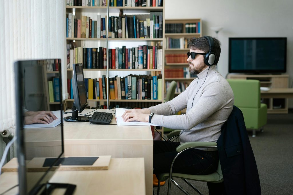
For Chapman, the results have been nothing short of transformative. He now wears dark glasses to reduce glare, but otherwise enjoys the freedom of sight. Everyday tasks that once required assistance, reading signs, crossing streets, and recognizing faces are now within his grasp. He dreams of returning to basketball, traveling to new places, and experiencing the visual world he missed for so long.
His journey has also had a profound emotional impact. After decades of blindness, making eye contact with another person was overwhelming. “It’s like getting my life back,” Chapman said. Beyond the practical benefits, regaining vision has given him back a sense of connection to others, the world, and himself. It is a new chapter one he once thought impossible.
Chapman’s story also inspires hope for others with similar conditions. By sharing his experience publicly, he shines a light on an option that many may not even know exists. His courage in undergoing such a radical surgery has opened the door for others who may one day see again thanks to this procedure.
Science, Resilience, and the Unlikely Power of a Tooth
Brent Chapman’s story goes beyond medicine. It reminds us how science can follow strange and ingenious paths. A tooth, designed to chew food, became a vessel for sight. A surgeon’s persistence and a patient’s resilience turned an outlandish idea into a life-changing reality. It shows how solutions to some of humanity’s most difficult problems can emerge from the most unexpected sources.
For those living with blindness, Chapman’s success is a beacon of hope. For the rest of us, it’s an invitation to marvel at the creativity of medicine and the resilience of the human spirit. The story of a man who regained sight through his own tooth shows science at its most human. And as surgeons refine and expand this procedure, the future of vision restoration may hold even more astonishing possibilities, reminding us that sometimes, the key to unlocking life’s greatest challenges lies in the unlikeliest of places.