Your cart is currently empty!
Doctors Are Destroying Tumors Inside MRI Scanners Without Making a Single Incision

Australia has quietly achieved something that sounds like it belongs in the future of medicine. At a hospital in Sydney, doctors are now destroying certain cancer tumors by freezing them from the inside while watching the entire procedure unfold through an MRI scanner. There are no large surgical cuts, no operating-room incisions, and in some cases, patients are returning home within a day. For people facing the prospect of invasive cancer surgery, the development offers a glimpse at a very different path forward.
Contents
show
The technology is already being used on real patients at Liverpool Hospital, where medical teams have combined advanced MRI imaging with a procedure known as cryoablation. The result is a treatment that allows doctors to target tumors with extraordinary precision while minimizing damage to healthy tissue around them. One patient who had been struggling with severe pain from a spinal tumor reported being pain-free within 24 hours of treatment, drawing attention to a breakthrough that could reshape cancer care in Australia and beyond.
Inside The MRI Machine That’s Freezing Tumors To Death
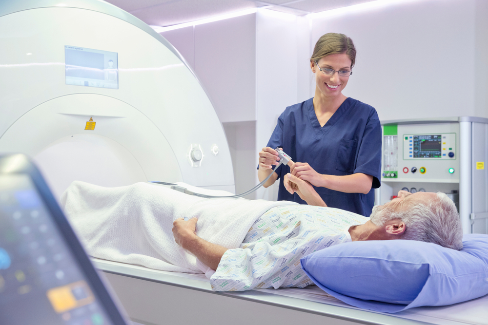
The procedure is known as MRI-guided cryoablation. While cryoablation itself has been used for years in medicine, performing it under continuous MRI guidance represents a major step forward in precision treatment. During the procedure, doctors insert a thin needle-like device called a cryoprobe directly into or alongside a tumor through a tiny puncture in the skin.
Once positioned, the cryoprobe rapidly cools to extremely low temperatures. Ice crystals begin forming inside the cancer cells, damaging their internal structures and cutting off blood flow. As the freezing process continues, the tumor tissue dies while nearby healthy tissue is preserved as much as possible.
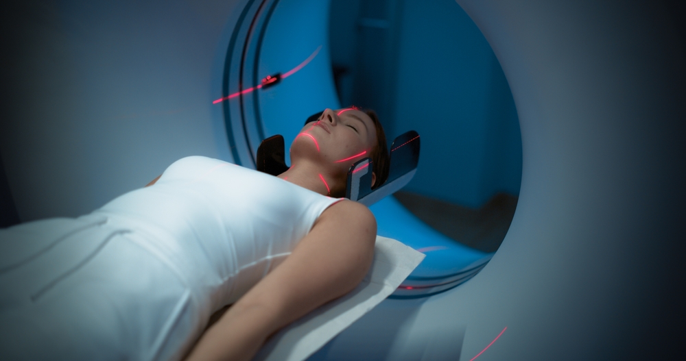
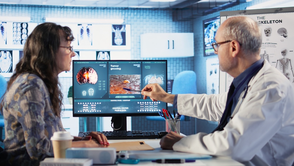
The real advantage comes from the MRI scanner itself. Doctors can see detailed images of the tumor, surrounding nerves, blood vessels, and organs throughout the entire procedure. Instead of relying on estimates, they can monitor exactly where the probe is located and how the treatment is progressing in real time.
An expanding frozen area known as an “ice-ball” develops around the tumor as treatment continues. Medical teams can watch that frozen zone grow on the MRI screen and make adjustments whenever necessary. Dr. Glenn Schlaphoff described the process simply: “That ice is used to kill the tumour in a very neat, discreet way.”
A Grandmother Went From Agony To Relief In Just One Day
One of the first people to undergo the procedure was 63-year-old Sydney grandmother Josephine Cordina, who had been living with severe pain caused by a small tumor lodged in her spine. Everyday activities had become increasingly difficult, and finding a comfortable position to sit or lie down was almost impossible.
Traditional surgery would have involved a far more invasive approach. Doctors would likely have needed to stabilize her spine using screws and other hardware, followed by a lengthy recovery period that could have lasted weeks or months.
Instead, she became one of the first patients to receive MRI-guided cryoablation. Rather than opening the body through major surgery, doctors used the cryoprobe to target the tumor directly while monitoring the treatment through MRI imaging.
The results were dramatic. Within 24 hours, her pain was gone. Instead of recovering from a major operation, she returned home with little more than a small puncture mark where the probe had entered her body.

Why Doctors Believe This Could Change Cancer Treatment
For decades, cancer patients have often faced difficult choices between effective treatment and the physical toll associated with surgery. Major operations can involve significant pain, lengthy hospital stays, visible scarring, and extended rehabilitation.
MRI-guided cryoablation offers a different approach. Because the procedure requires only a small entry point for the cryoprobe, there is no need for large incisions. Muscles remain largely undisturbed, and surrounding tissue experiences far less trauma than it would during conventional surgery.
Recovery times can be dramatically shorter as a result. Some patients leave the hospital on the same day, while others return home the following morning. Reduced recovery times also help free up hospital beds and medical resources that can be used for other patients.
The technology may also expand treatment options for people who were previously considered poor candidates for surgery. Older patients, individuals with underlying health conditions, and those with tumors located in difficult areas may benefit from a less invasive alternative.
The Technology Could Soon Be Used On More Types Of Cancer
Although spine tumors have been among the first conditions treated using the new system, doctors are already exploring additional applications. Researchers believe the same approach could potentially be used for a wide range of cancers throughout the body.
Liver tumors are one of the most promising targets. Surgery involving the liver can be complex due to the organ’s structure and blood supply. A highly targeted freezing technique could offer doctors another option for selected patients.
Kidney tumors are also attracting interest. Because cryoablation focuses treatment on a specific area, it may allow doctors to destroy cancerous tissue while preserving more healthy kidney function than traditional surgery in some cases.
Soft tissue tumors located in muscles and connective tissues are being studied as well. Researchers are also examining whether cryoablation could be combined with immunotherapy treatments and targeted cancer drugs to improve outcomes and reduce the risk of recurrence.
What Makes MRI Better Than Other Imaging Methods?
Doctors have several tools available when performing minimally invasive procedures, including CT scans and ultrasound imaging. Each has strengths, but MRI offers unique advantages when paired with cryoablation.
One of the most important benefits is image quality. MRI scanners provide exceptional detail when visualizing soft tissue structures. This becomes particularly important when tumors are located close to nerves, blood vessels, or vital organs.
MRI also allows doctors to see the frozen treatment zone as it develops. During cryoablation, physicians can monitor the exact size and shape of the ice-ball surrounding the tumor. If adjustments are needed, they can make them immediately rather than discovering a problem afterward.
Another advantage is the absence of radiation exposure. Unlike CT imaging, MRI relies on magnetic fields and radio waves. Patients who require multiple scans over time can therefore avoid additional radiation exposure during treatment.
These advantages combine to create a level of precision that many cancer specialists believe represents an important step forward in minimally invasive oncology.
There Are Still Challenges Ahead
Despite the excitement surrounding the technology, doctors stress that MRI-guided cryoablation is not suitable for every patient or every tumor. Careful evaluation remains essential before treatment decisions are made.
Tumor size remains an important factor. Very large cancers may still require traditional surgery or other forms of treatment. Location also matters, particularly when tumors are positioned near critical structures that cannot safely tolerate freezing.
Another question involves long-term outcomes. Because the technology is relatively new, researchers are continuing to monitor patients to determine how results compare with conventional surgery over many years.
Cost presents another challenge. MRI scanners, cryoablation equipment, and specialist treatment teams require substantial investment. While larger medical centers may be able to adopt the technology quickly, smaller hospitals could face greater barriers.
Australia’s Breakthrough Could Influence Cancer Care Worldwide
Medical centers around the world are already watching Australia’s experience closely. If the technology continues producing strong results, experts believe it could help reduce healthcare costs by shortening hospital stays and lowering complication rates.
The approach also fits into a broader shift toward more personalized cancer treatment. Rather than removing large areas of tissue, doctors can focus precisely on the problem area while preserving as much healthy tissue as possible.
Liverpool Hospital’s team is already exploring ways to expand the program, including the possibility of robotic assistance that could make probe placement even more accurate. Researchers are also working on protocols that would allow more patients to receive treatment and return home on the same day.
Cancer treatment has evolved dramatically over the past several decades, but many therapies still rely on methods that can be physically demanding for patients. Australia’s new MRI-guided system points toward a future where destroying a tumor may require nothing more than a precisely placed probe, an MRI scanner, and a carefully controlled burst of extreme cold.
Sources:
- ASMIRT. (2026c, March 3). New MRI-Guided Cryoablation Advances Treatment Options – ASMIRT. ASMIRT -. https://asmirt.org/news/new-mri-guided-cryoablation-advances-treatment-options/
- MRI guided cryoablation — Spectrum Interventional radiology. (n.d.). Spectrum Interventional Radiology. https://www.spectrumir.com.au/mri-cryoablation?
Australia, cancer care, cancer patients, Cancer Research, cancer treatment, Cryoablation, future medicine, health news, healthcare innovation, Liverpool Hospital, medical breakthrough, medical innovation, Medical Technology, MRI Technology, oncology, scientific discovery, Spine Tumor, Sydney News, Tumor Treatment