
Your cart is currently empty!
Scientists Find New Ways to Make Cancer Cells Destroy Themselves
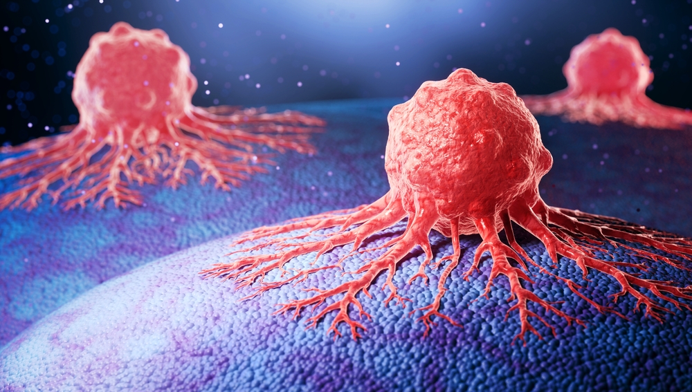
Cancer cells have developed ways to survive conditions that would normally damage or destroy them. Now, researchers are investigating several ways to exploit those vulnerabilities, with experiments revealing how physical pressure, targeted protein inhibition and tiny nanoparticles can trigger cancer cell death.
Contents
show
The findings come from separate research projects, but they point toward a fascinating possibility for future cancer treatment. Scientists are exploring whether the same biological systems that cancer cells use to survive could eventually become the mechanisms that cause their destruction.
Scientists Discover What Happens When Cancer Cells Are Trapped
Cancer cells face a difficult physical challenge when they spread beyond their original tumor. To invade surrounding tissues and potentially reach other parts of the body, they must squeeze through narrow spaces while adapting their shape and movement.
Researchers from Fudan University, Sun Yat-sen University and the Chinese Academy of Sciences in Shanghai investigated what happens when cancer cells remain trapped under physical pressure for an extended period. Their experiments used three-dimensional models designed to reproduce some of the physical challenges cells encounter during metastasis.
The researchers gently compressed cancer cells into extremely thin spaces while preventing them from attaching to surrounding surfaces. They maintained these conditions for several hours, allowing them to observe how the cells responded to prolonged mechanical stress.
Study authors Liming Sun and Yan-Jun Liu told Newsweek that the experimental system allowed them to follow the cells as they attempted to escape their confined surroundings. The researchers observed that the cells initially switched into a fast-moving, amoeba-like state before eventually losing their ability to move.
Sun and Liu described an unexpected discovery in which prolonged confinement caused the cells to stop moving and activate a built-in cell death program. The results suggested that sustained physical stress could push some cancer cells toward a biological process that ends in their destruction.
The team also found that the cells lost their polarized shape before dying. This change effectively stopped their invasive behavior, suggesting that the physical conditions affected their ability to spread before the cell death process took hold.
How Physical Pressure Activates a Cell Death Mechanism
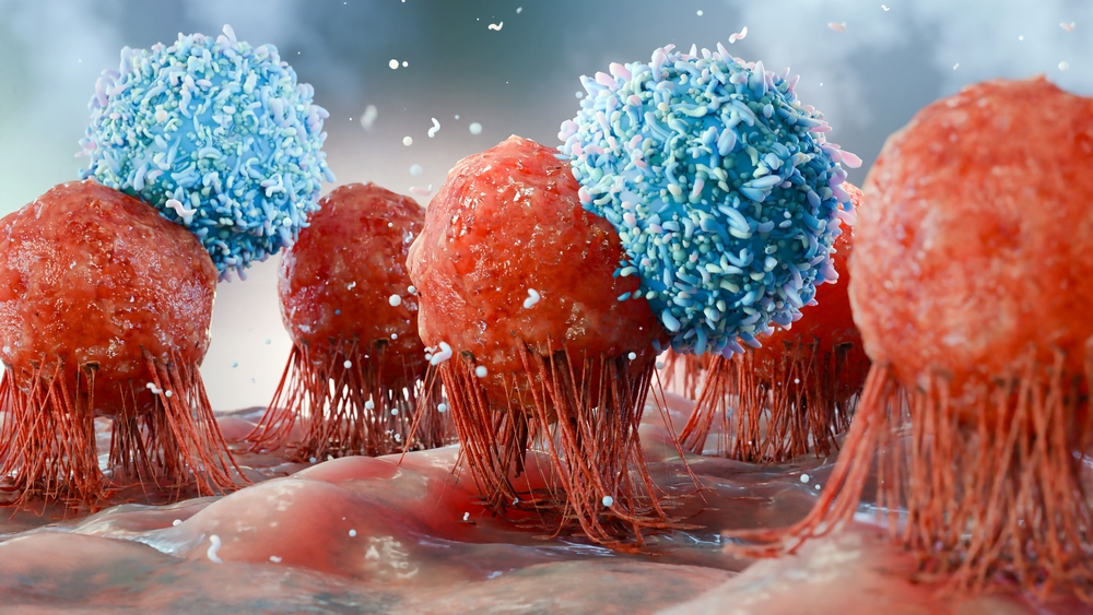
The Shanghai researchers focused on a biological process known as the mesenchymal-to-amoeboid transition, or MAT. During this transition, cells can rapidly change their shape and movement, helping them adapt to tight spaces without requiring new genetic transcription.
This flexibility can help cancer cells overcome physical obstacles as they move through surrounding tissues. However, the researchers found that prolonged confinement could turn that adaptation into a disadvantage.
The experiments showed that sustained mechanical stress eventually activated MLKL, short for mixed lineage kinase domain-like. This protein plays a role in necroptosis, a regulated form of cell death that can eliminate cells under particular conditions.
The researchers suggested that cancer cells may initially respond to confinement by attempting to escape, only to encounter stress that becomes increasingly difficult to tolerate. When that stress persists, the biological response can shift from adaptation toward cell death.
Their findings also revealed a potential complication. According to Sun and Liu, tumors may gradually become enriched with cells expressing lower levels of MLKL, which are more resistant to necroptosis and may possess greater migratory and invasive abilities.
This suggests that the same pressure capable of eliminating vulnerable cancer cells could also favor the survival of more resistant populations. Further research will be needed to determine how this mechanism operates across different cancers and whether it can be exploited safely in treatment.
Blocking One Protein Reduced Lung Tumor Growth by 80%

Another research team investigated whether cancer cells could be pushed toward destruction by blocking a protein that helps them suppress a different form of cell death. Researchers at NYU Langone Health focused on ferroptosis, a process driven by iron-dependent damage to cellular membranes.
Cancer cells can develop defenses against ferroptosis, allowing them to survive despite the damaging conditions associated with rapid growth. The NYU researchers examined whether disabling one of these defenses could make lung cancer cells more vulnerable.
Their study, published in Nature, investigated ferroptosis suppressor protein 1, commonly known as FSP1. Researchers found that blocking this protein reduced tumor growth by up to 80% in mice with lung adenocarcinoma, a common form of lung cancer.
The team genetically engineered mice so that their lung cancer cells lacked the FSP1 gene. These animals developed significantly smaller tumors as cancer cell death increased, suggesting that FSP1 helped the cancer cells resist the process.
Researchers also tested an experimental inhibitor called icFSP1. Treatment improved overall survival among mice carrying lung tumors, producing results approaching those seen in mice whose cancer cells had been genetically engineered to lack FSP1.
Senior study author Thales Y. Papagiannakopoulos, an associate professor at NYU Grossman School of Medicine, said: “This first test of a drug that blocks ferroptosis suppression highlights the importance of the process to cancer cell survival and paves the way for a new treatment strategy.”
The findings offer a potential direction for developing future cancer therapies. However, the reported tumor reduction occurred in mice, and the study does not establish that icFSP1 is safe or effective for treating human patients.
What Ferroptosis Does Inside Cancer Cells

Ferroptosis differs from the cell death process investigated in the mechanical confinement research. Rather than being triggered by prolonged physical pressure and MLKL activation, it involves iron-dependent chemical reactions that damage cells.
Iron can contribute to the production of reactive oxygen species, highly reactive molecules that form during chemical reactions involving oxygen. These molecules play roles in normal cellular signaling, but excessive amounts can cause oxidative stress and damage important cellular components.
When reactive oxygen species accumulate, they can attack fats in cell membranes and disrupt the structures cells need to survive. Ferroptosis occurs when this iron-dependent damage becomes sufficient to trigger cell death.
Cancer cells can counter this threat by activating protective mechanisms. FSP1 is one of the proteins involved in suppressing ferroptosis, making it a potential target for treatments designed to restore the cell death process.
The NYU researchers also investigated glutathione peroxidase 4, or GPX4, another protein involved in preventing ferroptosis. Their findings suggested that FSP1 could be a more selective target because it played a greater role in blocking ferroptosis in lung cancer cells while having a smaller role in normal cellular functions.
That difference could potentially translate into fewer side effects, although this possibility requires further investigation. The researchers also found that higher FSP1 levels were associated with poorer survival among human patients with lung adenocarcinoma, providing another reason to investigate the protein’s role in the disease.
Tiny Silica Nanoparticles Trigger Another Wave of Cell Death

A separate collaboration between researchers at Memorial Sloan Kettering Cancer Center and Cornell University explored whether tiny silica nanoparticles could trigger ferroptosis without carrying conventional cancer-killing drugs.
The particles, known as C dots, are extremely small silica structures containing fluorescent dye molecules. They can glow under particular light conditions, allowing researchers to track their movement through the body and investigate their potential diagnostic applications.
Researchers discovered that these nanoparticles could also induce ferroptosis under the right conditions. Their experiments showed that the particles triggered cancer cell death across several cancer cell types, while tests in cancer-bearing mice demonstrated significantly smaller tumors and inhibited tumor growth.
The team also found that the lethal effect could be reversed by administering a drug that inhibits ferroptosis. This supported the researchers’ conclusion that the cell death process played a central role in the observed results.
Study leader Michelle Bradbury, a clinician-scientist at Memorial Sloan Kettering, said: “This is the first time we have shown that the particle has intrinsic therapeutic properties without associated toxicity.”
The discovery could help researchers investigate treatments that use the physical properties of nanoparticles rather than relying exclusively on toxic drugs attached to their surfaces. Conventional approaches have often involved loading nanoparticles with therapeutic compounds intended to kill cancer cells.
C dots had previously demonstrated potential in diagnostic applications, and the research reference reports their use in clinical settings involving melanoma. However, their ability to induce ferroptosis represents a separate therapeutic question that requires additional investigation.
The research team is examining which cancers are most sensitive to the particles and whether the approach could work alongside established treatments. Scientists are also investigating whether the particles could influence immune cells and the tumor microenvironment to support additional anticancer activity.
Three Research Approaches Target Cancer Cell Survival

Although the studies investigated different biological mechanisms, they share a common research question: can scientists exploit vulnerabilities that cancer cells depend on to survive and spread?
The approaches described in the research include three distinct strategies, each with its own potential advantages and unanswered questions.
- Mechanical confinement: Researchers found that prolonged physical pressure activated an MLKL-associated cell death pathway in experimental cancer cell models. The findings also showed that resistant cells could survive and potentially become more invasive.
- FSP1 inhibition: NYU researchers used an experimental inhibitor to suppress a protein that protects cancer cells against ferroptosis. The approach reduced lung tumor growth by up to 80% in mice, although its safety and effectiveness in people remain unestablished.
- Silica nanoparticles: Memorial Sloan Kettering and Cornell researchers found that C dots could trigger ferroptosis in several cancer cell types. Their experiments also demonstrated reduced tumor growth in cancer-bearing mice.
These approaches come from separate research projects and have not been established as a single combined treatment. Each targets a different vulnerability, meaning researchers will need to determine which strategy is suitable for particular cancer types and circumstances.
The studies also demonstrate why cancer treatment research requires careful evaluation. A process that destroys one group of cancer cells may leave behind another population that is better equipped to survive, while an experimental drug or nanoparticle may behave differently in a living person than in a laboratory model.
Researchers Are Exploring the Next Steps

The next phase of research will involve determining how these mechanisms behave across different tumors and whether they can be translated into effective treatments.
The NYU team plans to optimize FSP1 inhibitors and investigate whether ferroptosis could be targeted in other solid tumors, including pancreatic cancer. Lead study author Katherine Wu, an MD/PhD student working in the Papagiannakopoulos laboratory, said the researchers aim to translate their findings into potential clinical therapies.
The nanoparticle research team is also investigating how C dots might be combined with existing cancer treatments. Researchers are evaluating which cancers are most susceptible to the particles and whether they could influence immune cells and the surrounding tumor environment.
For the mechanical confinement research, a key challenge will be understanding why some cancer cells die while others survive and potentially become more aggressive. The researchers also need to establish whether the observed responses can be reproduced in more complex biological systems.
Human clinical trials will be essential for determining whether these experimental approaches can benefit patients. Such studies must establish appropriate treatment strategies, identify potential risks and determine whether the benefits outweigh the harms.
What These Findings Mean for Future Cancer Treatment
The findings offer several promising research directions, but they do not establish a new cure for cancer. The 80% reduction in tumor growth reported in the FSP1 experiments occurred in mice, while the mechanical confinement findings came from experimental models and the nanoparticle research included laboratory and animal studies.
The distinction is important because results in early experiments do not automatically translate into safe and effective treatments for people. Cancer is also a collection of different diseases, and a strategy that works against one tumor type may not produce the same response in another.
For now, researchers are gaining a better understanding of how cancer cells protect themselves against biological stress and how those defenses might be disrupted. By investigating the mechanisms that allow tumors to survive, scientists hope to identify new ways to limit cancer growth and prevent its spread.
The central challenge is turning these discoveries into treatments that can reliably distinguish vulnerable cancer cells from healthy tissue. If researchers can overcome that hurdle, the biological defenses cancer cells rely on today could eventually become targets for tomorrow’s therapies.